Peripheral Arterial Disease
Peripheral Arterial Disease: Causes, Symptoms, and Treatment
Peripheral Arterial Disease (PAD) is a narrowing of the peripheral arteries serving the outer regions of the body, including the legs, stomach, arms and head, caused by a buildup of cholesterol and fatty deposits (plaque) which narrows or blocks blood flow to these arteries. The supply of oxygen to cells is also limited due to the plaque buildup in the artery walls. PAD most commonly affects arteries in the legs.
Symptoms of PAD include:
Insurance participation varies by provider. Please call our Arizona Heart and Vascular office to verify insurance participation and to understand co-payment and referral requirements for your insurance plan.
- Heaviness, cramping, pain or fatigue in the leg or hip muscles on exertion. Typically, this pain goes away with rest and returns with activity.
- Lack of growth of your toenails and leg hair
- One foot feeling colder than the other
- Pale, discolored, or blue leg or foot
- Leg weakness or numbness
- Pain or a feeling of pins and needles in your extremity
- Pain at rest in your leg and foot
- Sores or wounds on your extremities that heal slowly or not at all. The sores may become infected.
PAD affects more than 18 million people
More than cancer, stroke, and congestive heart failure. 1 in 3 people over the age of 50 who suffer from diabetes also have PAD. Early detection is important. If you think you might be at risk for the disease, consult a health care professional.
Risk Factors
- Diabetes
- Family history
- High Blood Pressure / Hypertension
- Smoking
- Obesity / Unhealthy Diet
- High Cholesterol
- Kidney Disease
- Lack of exercise
- Stress
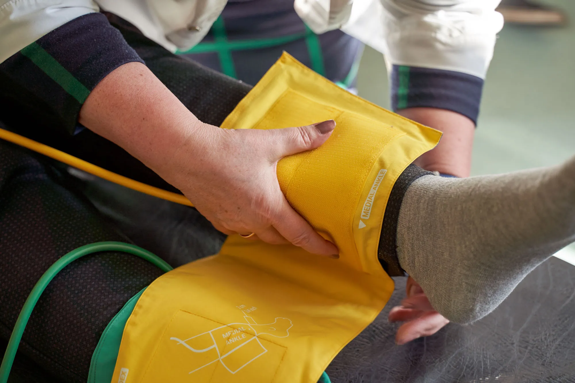
ABI Testing – Detecting Peripheral Artery Disease
An Ankle Brachial Index (ABI) is a simple test that a doctor can perform in the office to help determine if you have PAD. The test compares the blood pressure in your ankle to that in your arm, if the pressure is lower in your ankle than your arm, there is a chance you have PAD.
Upper / Lower Extremity Doppler
This test uses ultrasound to look at the blood flow in the large arteries and veins in the arms or legs. During the ultrasound, the technician places a handheld instrument called a transducer on your skin, transmitting sound waves that produce images of the blood vessels on a monitor. This test is done as the first step to look at arteries and veins. Sometimes, arteriography and venography may be needed later. The test is done to diagnose:
- Arteriosclerosis of the arms or legs
- Blood clot (deep vein thrombosis)
- Venous insufficiency
- Injury to the arteries
- Monitor arterial reconstruction and bypass grafts
Peripheral Angiography
In an angiogram, X-rays are used to determine whether plaque has blocked the blood vessels. During this test, which requires local anesthesia, a doctor makes a small incision in the skin near the groin, inserts a thin tube called a catheter, and guides it into an artery. The doctor moves the catheter to the area to be examined and injects a dye that highlights any narrow, enlarged, or blocked blood vessels.
Lower Extremity Angioplasty & Stenting
Angioplasty involves passing a catheter with a balloon through a blocked artery. Once inflated, the balloon compresses the plaque against the wall of the artery. During angioplasty, a tiny metal mesh tube called a stent may be placed in the artery to help hold it open.
Atherectomy
Atherectomy uses a special catheter to gently shave and remove plaque from the arteries.
Endovascular Aortic Aneursym Repair
An aortic aneurysm is a bulging, dilation or ballooning in the wall of the aorta. The aneurysm occurs when a weakness develops in a portion of the artery wall. As the aneurysm enlarges, IT stretches the walls of the artery thinner which compromises the artery wall’s ability to stretch any further, like a fully inflated balloon. Just as a balloon will pop when blown up too much, an aneurysm is at risk of rupturing and causing potentially fatal bleeding. Endovascular aneurysm repair involves inserting a graft within the aneurysm through small groin incisions using X-rays to guide the graft into place.